Age-related androgen deficiency syndrome in men

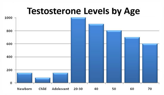
The syndrome of age-related androgen deficiency in men is a violation of the biochemical balance that occurs in adulthood due to a lack of androgens in the blood serum, often accompanied by a decrease in the body’s sensitivity to androgens. As a rule, this leads to a significant deterioration in the quality of life and adversely affects the functions of almost all body systems. Naturally, the issues of androgen deficiency therapy are of great interest, since it poses a difficult task for a clinician: to choose from a wide arsenal of methods and drugs of hormone therapy the most optimal, combining quality, efficiency, and ease of use.

Currently, urologists and andrologists most often use testosterone replacement therapy. This method allows you to solve a number of tasks: to reduce the symptoms of age-related androgen deficiency by increasing libido, overall sexual satisfaction, to reduce the severity or completely eliminate vegetative-vascular and mental disorders. In addition, if testosterone replacement therapy is used for more than 1 year, patients experience an increase in bone density, a decrease in the severity of visceral obesity, as well as an increase in muscle mass. Also, after a long course of treatment, laboratory parameters are normalized: there is an increase in the level of hemoglobin or the number of red blood cells, a decrease in the level of VLDL (very low-density lipoproteins) and LDL (low-density lipoproteins) with an unchanged level of HDL (high-density lipoproteins). Many authors believe that such an effect can be achieved by restoring the concentration of testosterone in the blood to a normal level (10-35 nmol/l). It should also be taken into account that * 17α-alkylated testosterone preparations fluoxymesterolone and methyltestosterone have pronounced hepatotoxicity, having a toxic and carcinogenic effect on the liver, and also negatively affect the blood lipid spectrum (a sharp increase in the level of atherogenic and a decrease in the level of anti-atherogenic lipoproteins). Therefore, the use of these testosterone derivatives in clinical practice was discontinued.

Currently, testosterone undecanoate is preferred among oral medications. This testosterone ester is not subjected to primary hepatic metabolism, since it is absorbed into the lymphatic system, bypassing the liver. After the hydrolysis of testosterone undecanoate in the lymphatic system, testosterone enters the systemic bloodstream, which has a therapeutic effect both by itself and through its main metabolites-dihydrotestosterone (DHT) and estradiol, which cause the full spectrum of androgenic activity of testosterone. Thus, testosterone undecanoate retains its activity when administered orally. At the same time, bypassing the portal vein system and passing through the liver, testosterone undecanoate does not have hepatotoxic and hepatocancerogenic effects. The half-life of the drug from the plasma is 3-4 hours. In this regard, the dosage regimen of testosterone undecanoate is a 2-fold intake during the day, this is not always convenient for patients. Based on our own experience, we believe that Andriol is a fairly mild drug and helps only in cases of initial and minimal manifestations of age-related androgen deficiency.
androgen deficiency

Intramuscular injections of prolonged testosterone esters are also a widely used method of substitution therapy in men with hypogonadism. The two most well – known esters of testosterone, testosterone cypionate and testosterone enanthate, have similar pharmacokinetics. With intramuscular administration of these drugs, a depot is created from which the drug is released into the bloodstream. During the first 2-3 days after administration, the testosterone level rises to supraphysiological figures, and then slowly decreases over the next 2 weeks to subnormal values. The positive side of these drugs is the duration of the therapeutic effect. Nevertheless, sharp changes in the level of testosterone, often felt by the patient himself in the form of rises and decreases in libido, general well-being, emotional status, are undesirable qualities of these drugs. In this regard, great hopes are pinned on the new drug Nebido (Sharing), the pharmacokinetics of which is significantly different from other testosterone esters. Nebido is a testosterone undecanoate and is a drug that does not have a peak increase in concentration.

Over the past two decades, much attention has been paid to the study of the benefits of transdermal use of testosterone preparations. Scrotal patches have an effective effect, and some patients consider them the most convenient method of treatment. Skin patches are most well perceived by patients and give an effective level of testosterone in the blood serum. Nevertheless, there are some differences between these two types of patches regarding their allergogenic potential: when using skin patches, there is a much higher frequency of allergic reactions and skin irritation than when using scrotal patches. Testosterone gel has all the advantages of patches and does not cause the development of skin reactions. Its only drawback is the possibility of contact of the gel with a partner and an insufficient number of long-term studies on its use.

The transdermal route of testosterone administration allows avoiding its primary metabolism in the liver and inactivation, as it happens when using oral androgenic drugs, and also allows simulating the circadian rhythms of the release of physiological unmodified testosterone and its natural metabolites, estradiol and DHT. In addition, therapy with the use of patches and gel can be easily interrupted if necessary. The positive aspects of this method of treatment also include a low risk of drug dependence.

Although the European drug 5-α-dihydrotestosterone gel (DHT) is recognized as effective, it is not known whether the isolated use of a non-aromatized androgen, such as DHT, has the same effect as testosterone, due to the fact that testosterone metabolites include estradiol. According to many authors, the use of the drug is not recommended, since DHT, due to the inability to convert into estradiol, does not have the full range of therapeutic properties of testosterone (for example, the effect on bone tissue and the cardiovascular system).

Thus, we can say that there is no optimal remedy for the treatment of age-related androgen deficiency in men. And the choice of the drug should be approached strictly individually, taking into account the patient’s age, body mass index, the need to preserve spermatogenesis, hematocrit indicators and concomitant diseases.

Female and male infertility: part 2

Male infertility: The maturation of spermatozoa in the testicles occurs under the influence of gonadotropins secreted by the pituitary gland, and male sex hormones (androgens). Unlike the female body, there is no cyclicity in the regulation of the male reproductive system.

Diagnosis and treatment of male infertility

The sexual potency of a man is not an indicator of his fertility, since the ability to fertilize is determined by the quality of spermatozoa. In men, in addition to determining the level of male sex hormones and their daily rhythm, it is necessary to examine the spermogram before, during and after treatment. Male fertility is determined by the following key factors: spermatogenesis, sperm transport, the ability of the sperm to penetrate into the egg to introduce genetic material into it.

To identify the causes of infertility, it is important to know the following features of sperm transport. The seminal fluid forms a gel almost immediately after ejaculation, but after 20-30 minutes it is liquefied under the influence of enzymes coming from the prostate gland. In some cases, the seminal fluid remains viscous, retains spermatozoa and prevents them from moving into the uterus. Normally, the acidity of the seminal fluid is higher than 6 pH, which protects the spermatozoa from the harmful effects of the acidic pH of the vagina.

Male infertility

On the way from the vagina to the tubes, the number of spermatozoa decreases significantly. On average, out of 200-300 million spermatozoa that have entered the vagina, eggs reach less than 200. Some of the spermatozoa are destroyed under the influence of vaginal enzymes, as well as as a result of “squeezing” the seminal fluid from the entrance to the vagina. The largest number of spermatozoa dies during passage through the fallopian tubes. Phagocytosis of spermatozoa occurs throughout the reproductive tract.

Examination and treatment of patients to determine the cause of infertility is carried out by an andrologist or urologist.

Examination of a man with infertility begins with the analysis of a spermogram, which allows you to determine the volume of sperm, the total number of spermatozoa, the number of active and normal spermatozoa. In addition, the number of white blood cells is calculated, the viscosity of the sperm is determined, the dilution time is determined. The lack of dilution of seminal fluid is one of the causes of male infertility.

Another factor leading to male infertility may be sperm agglutination. At times, this happens in most men. However, the detection of sperm agglutination in repeated samples indicates an autoimmune reaction or infection. If a large number of abnormal spermatozoa are detected, an additional study is shown – a morphological analysis of sperm. Based on the conducted studies, the nature of the pathology of spermatozoa is established and the method of infertility treatment is determined. The maturation of spermatozoa largely depends on the impact of internal and external adverse factors. Alcohol, nicotine, occupational hazards, stress, nervous and general fatigue, acute and chronic diseases worsen the quality of sperm.

The composition of spermatozoa is resumed within three months. This should be taken into account when submitting sperm for analysis. The longer it is possible to maintain a healthy lifestyle, the better the result. Before submitting sperm for analysis, it is necessary to refrain from sexual contact for 3-5 days. Compliance with this condition allows you to get the best composition and quality of spermatozoa. Daily or more frequent ejaculations can lead to a decrease in the quality of spermatozoa, but abstinence for 5-7 days or longer is also undesirable, since an increase in the number of spermatozoa does not contribute to the “preservation of sperm” (as some patients believe), but is accompanied by a decrease in their mobility as a result of an increase in the proportion of old cells.

The most common causes of male infertility are:

  • inflammatory diseases (prostatitis, urethritis, etc.);
  • obstruction (obturation) of the vas deferens;
  • dilation of the veins of the spermatic cord (varicocele);
  • hormonal and sexual disorders.

It is not always possible to identify all possible causes of infertility. The male factor of infertility occurs in 40% of cases; unidentified causes-in 10%.

During the examination, physical disorders may be detected, for example, pronounced hypospadias, in which sperm does not enter the vagina. It is extremely rare for diabetes mellitus, neurological disorders, after prostatectomy, retrograde ejaculation of sperm into the bladder can occur.

In some cases, to find out the cause of infertility, in addition to a spermogram, a testicular biopsy should be performed. Azoospermia (the absence of spermatozoa in the sperm fluid) in the presence of spermatogenesis indicates an obstruction of the ducts. If a biopsy reveals complete hyalinization and fibrosis of the seminal tubules, the possibility of fertility is almost excluded.

The most common cause of male infertility is inflammatory processes directly in the testicles (orchitis, orchiepididymitis) and in the vas deferens (epididymitis, differentitis, vesiculitis). Approximately 15-20% of men who have suffered from mumps have orchitis, which in half of the cases ends with hypoplasia of the testicles of various degrees. If an infection of the genitourinary tract is detected, antibacterial therapy and, possibly, prostate massage should be prescribed.

25% of infertile men have varicose veins of the left internal seminal vein, the ligation of which in 50% of cases determines the possibility of fertilization. With clinically expressed varicocele, the size of the left testicle is usually reduced. It should be noted that varicocele is detected in 10-15% of men in the general population, and about half of them have violations of the spermogram. Even a weakly expressed varicocele can affect the quality of sperm, which is associated with an increase in the temperature of the testicles. Men with a normal spermogram, but who have been diagnosed with varicocele, need periodic examination, because their spermogram indicators may worsen over time.

Endocrine disorders rarely cause male infertility. Nevertheless, it is necessary to study the content of FSH, LH, testosterone and prolactin. If an increased level of prolactin is detected, a further diagnostic examination is performed to identify/exclude a pituitary tumor.

There are cases of infertile marriage, when both sexual partners are healthy, and pregnancy does not occur. This is often due to their immunological incompatibility, when antibodies to spermatozoa are produced in a woman’s body. These proteins inhibit the motility of spermatozoa and their ability to fertilize an egg.

There are special methods for diagnosing immunological incompatibility. The simplest of them – the postcoital test-allows you to get information about the absorbing ability of cervical mucus, as well as about the ability of spermatozoa to reach it and survive in it. The test is planned for the expected day of ovulation, which is determined by the previous basal temperature maps, the duration of previous cycles and the degree of humidity of the vagina. The cervical mucus is taken no later than 12 hours after coitus with the previous 48-hour abstinence; the cervical mucus is taken with tweezers. The extensibility of the mucus should be at least 8-10 cm. The mucus obtained during ovulation contains 90-95% water; it should be watery, liquid, transparent and abundant, and also should not contain cells. When drying on a slide, the mucus resembles a fern leaf. Mucus before ovulation and starting from 24-48 hours after it has a thick and viscous consistency, dries out in the form of amorphous lumps. The mucus of the optimal composition for the survival of spermatozoa usually persists for 2-3 days, but it varies from 1 to 5 days and even longer for different women. The normal number detected by a postcoital test is considered to be from 1 to 20 spermatozoa in the field of vision. If repeated tests do not detect spermatozoa or only find immobile cells, the prognosis is less favorable than when detecting live spermatozoa. Treatment becomes unsuccessful, especially in cases when repeated postcoital tests do not find spermatozoa or only dead forms are found, despite good mucus and a spermogram.

Male and female infertility

There are primary and secondary female infertility. Primary infertility is spoken of if a woman has not had a single pregnancy, despite regular sexual activity for a year in the absence of contraception, secondary – if there is a pregnancy earlier, that is, if it is impossible to conceive a child after an abortion, ectopic pregnancy, miscarriage, or the birth of a child. Voluntary infertility can be called a situation if pregnancy is undesirable and a woman is protected using contraceptives. There are also absolute infertility, when conception is impossible due to congenital pathology or irreversible changes in the woman’s body, and relative infertility, when the ability to fertilize is not impaired; in such cases, it is appropriate to talk about reduced fertility.

The inability of a mature woman’s body to conceive is due to the following reasons:

  • endocrine disorders – anovulation) – 40%;
  • tubal-peritoneal factor-30%;
  • gynecological diseases – 15-25%;
  • immunological factor – 3%;
  • unidentified factors-2-3%.

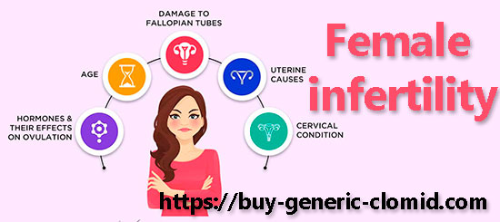
The main cause of ovarian infertility is anovulation – a violation of the menstrual cycle, as a result of which the maturation and release of the egg from the follicle does not occur. Endocrine (anovulatory) infertility can have different origins: hypothalamic, hypothalamic-pituitary, ovarian, thyroid and adrenal gland diseases, chromosomal abnormalities, violation of implantation of a fertilized egg, violation of the function of the fallopian tubes, etc.

Etiological factors of anovulatory infertility

Hypothalamic causes. Hypothalamic gonadoliberin deficiency (gonadotropin-releasing hormone-GnRH) leads to a violation of the regulation of the gonadotropic function of the pituitary gland and, accordingly, ovarian function; clinically manifested by anovulation. Violation of GnRH secretion can occur with emotional overstrain, weight loss, under the influence of medications.

Pituitary causes. Micro-and macroadenomas of the anterior pituitary lobe, prolactinomas can lead to pathological hyperprolactinemia. In all cases, if a pituitary adenoma is suspected, magnetic resonance imaging (MRI) is necessary. A slight increase in the level of prolactin, most often temporary, can occur with emotional overstrain.

Ovarian causes. There are primary and secondary ovarian insufficiency. The cause of primary ovarian insufficiency is ovarian pathology, secondary-a decrease in the secretion of GnRH in the hypothalamus or gonadotropin hormones in the adenohypophysis. Premature ovarian insufficiency may have a genetically determined, autoimmune, idiopathic, functional (weight loss, physical activity, medications) origin.

The most common cause of ovarian anovulation (female infertility) is polycystic ovary syndrome. Often, the syndrome of luteinization of a non-ovulated follicle is detected. This condition can be caused by some medications (prostaglandin synthetase inhibitors), it is often observed in endometriosis, stress, hyperandrogenism, hyperprolactinemia, inflammatory processes in the ovaries. The diagnosis is established on the basis of ultrasound (ultrasound) or laparoscopy data.

The thyroid gland and the adrenal glands. Hypothyroidism or hyperthyroidism is accompanied by a violation of the function of the pituitary gland, ovaries, which leads to anovulation. In Itsenko-Cushing’s disease and other cases of hypercortisolemia, elevated levels of testosterone and cortisol cause suppression of the gonadotropic function of the pituitary gland, as well as ovarian dysfunction, while secondary polycystic ovaries may develop.

Chromosomal abnormalities lead to amenorrhea and are also accompanied by infertility.

Violation of the implantation of a fertilized egg occurs as a result of a decrease in the level of progesterone; it can be caused by deformation of the uterine cavity with submucosal uterine fibroids.

Violation of the function of the fallopian tubes can be a consequence of inflammatory processes of the uterine appendages, which lead to a violation of the capture of the oocyte as a result of the formation of peritubar adhesions and damage to the fimbria, as well as damage to the epithelium of the tubes. The function of the fallopian tubes can be impaired with endometriosis, destructive appendicitis, suppuration after surgery on the pelvic organs or abdominal cavity.

Smoking, alcohol abuse, drug use, psychological factors, and adverse environmental effects can contribute to a decrease in fertility.

Female infertility: Diagnostics

To determine the cause of female infertility, it is necessary to study the hormonal status, conduct ultrasound of the pelvic organs, adrenal glands, analyze the physical, mental and social health of the patient and compare it with the course of the disease, as well as with the results of the treatment. The study of the quality of life allows us to optimize tactics and strategy in the treatment of women with various diseases of the reproductive system.

The following diagnostic tests are performed:

  • determination of functional changes in the ovaries and uterus;
  • detection of urogenital tract infection;
  • assessment of the state of the uterine cavity and patency of the fallopian tubes;
  • identification of an immunological conflict between spouses.

The most informative studies of the hormonal function of the ovaries are ultrasound and hormonal monitoring, supplemented by basal temperature measurement. In infertility, the examination of patients should begin with determining the level of luteinizing (LH) and follicle-stimulating (FSH) hormones in the blood. In the early follicular phase of the cycle, the level of FSH should be below 3-5 IU/l. Exceeding the standard values indicates that the biological age of the ovaries is older than the chronological age of the woman. The study of the FSH content should be carried out together with the determination of the level of estradiol, since at the concentration of estradiol above 250 pmol/l, the level of FSH decreases (by the negative feedback mechanism). To exclude polycystic ovary syndrome, the ratio of LH/FSH is additionally calculated.

To assess the state of carbohydrate metabolism, you should measure the level of glucose and insulin in the blood on an empty stomach. If necessary, a glucose tolerance test is performed. In the middle of the luteal phase of the cycle, the level of progesterone is determined (5-7 days after the basal temperature rises). The function of the thyroid gland and the level of blood prolactin are studied.

Recently, it is recommended to start the examination of patients with infertility with a study of the content of prolactin in the blood, since an increased level of prolactin is diagnosed in 20-25% of patients with infertility and various menstrual cycle disorders, and in 40-45% of them, macro – and micro-tumors of the pituitary gland are the cause of hyperprolactinemia.

Ultrasound of the pelvic organs is performed at the initial stages of examination of patients with infertility, and hysterosalpingography – in the follicular phase of the cycle.

Female infertility: Treatment of hormonal infertility

In the treatment of anovulatory infertility, clomiphene citrate or gonadotropin preparations prepared from the urine of pregnant or postmenopausal women, and in recent years – obtained by genetic engineering, are prescribed to restore fertility.

female infertility

Clomiphene citrate is an antiestrogenic drug that has the ability to bind estradiol receptors in all target organs, including the hypothalamus, at the site of GnRH synthesis, which causes an increase in the secretion of gonadotropins and especially FSH. An increase in the level of FSH stimulates the maturation of follicles in the ovaries and leads to an increase in the concentration of estradiol. By suppressing the regulatory effect of endogenous estrogens, clomiphene citrate blocks the normal feedback mechanism, which causes an increase in the frequency of cyclic GnRH secretion. The drug is prescribed from the 2nd day of the menstrual cycle for 25-50 mg or 100 mg for 5 days, and in the case of amenorrhea – from the 2nd day of the induced menstrual cycle. Patients with polycystic ovary syndrome have an increased sensitivity to drugs that stimulate ovulation, so such patients are prescribed clomiphene citrate at a dose of no more than 25-50 mg. The effectiveness of treatment is evaluated using ultrasound. From the 9th-10th day of the menstrual cycle, the diameter of the dominant follicle should be monitored, and on the 13th-14th day – the preovulatory follicle, which should have a size of 16-26 mm. If the disappearance or gradual decrease of the dominant follicle is recorded during ultrasound, ovulation has occurred. If three or more follicles with a size of 18-22 mm are detected according to ultrasound data, sexual contact should be avoided. The level of serum progesterone exceeding 20 nmol/l on the 21st day of the menstrual cycle indicates ovulation.

Currently, ovulation stimulation is recommended for no longer than 6 months, which is associated with an increased risk of ovarian cancer when taking clomiphene citrate for more than 12 months. In real practice, due to frequent dose adjustment, the drug is often used for a longer time.

Of the side effects of clomiphene citrate, hot flashes are most often noted, which occur in 10% of cases and disappear after stopping taking the drug. Sometimes, against the background of clomiphene citrate therapy, an increase in the size of the ovaries may occur. Rarely, patients note such undesirable phenomena as nausea, vomiting, depression, nervousness, fatigue, insomnia, headache, weight gain, pain in the mammary glands. When prescribing high doses of the drug to patients with polycystic ovary syndrome, the frequency of side effects increases. In the absence of the effect of the use of clomiphene citrate in such patients, laparoscopic diathermocoagulation of the ovaries or the appointment of gonadotropins is necessary.

We talked about female infertility today, we will write about male infertility in our next article, stay with us on this blog and recommend us to your friends and acquaintances. We remind you that you can also buy Clomid using our referral link, which is located in the blog header!

The mechanism of action of clomiphene and its effectiveness

Action of clomiphene: According to the hypothesis, clomiphene directly stimulates the hypothalamus-pituitary-ovary system, which in turn affects the gynothalamic-pituitary system. This hypothesis is confirmed by clinical data that have shown the possibility of increasing the release of estrogens after taking clomiphene without first increasing FSH.

This hypothesis is also confirmed in experimental studies. Thus, Smith and Doy showed that when clomiphene is added to a medium containing testosterone and placental microsomes, the production of estrogens from testosterone increases by 1.6 times. The Hammerstein data indicate the possibility of enhancing the synthesis of progesterone from acetate in the human yellow body (in vitro) with the addition of large amounts of clomiphene by activating the 3-b-ol-dehydrogenase enzyme system. At the same time, with an increase in the dose of clomiphene, the synthesis of progesterone is sharply inhibited.

According to Israel, both assumptions should be accepted as important working hypotheses; obviously, clomiphene has the possibility of a dual effect, which is confirmed by an increase in the excretion of both gonadotropins and estrogens in women with removed ovaries. This can be explained by enzyme changes in the biosynthesis of estrogens not only in the ovary, but also in the adrenal gland, which causes an increase in the synthesis of estradiol, which in turn causes the release of gonadotropins necessary for ovulation.

Currently, it is known that clomiphene increases the release of FSH and LH as a result of binding to estrogen-dependent receptors of the hypothalamus, which causes the development of a follicle in the ovary and the appearance of an estradiol peak preceding the ovulatory peak of gonadotropins. At the same time, estrogens increase the sensitivity of the pituitary gland to Gn-RH and the ovaries to gonadotropins, which leads to ovulation. A partially local effect of clomiphene on the ovary is also possible.

Action of clomiphene

Thus, clomiphene performs its biological effect mainly due to its anti-estrogenic effect, which activates the gonadotropic function of the pituitary gland. Clomiphene has found wide application for the treatment of patients with endocrine forms of infertility due to its ability to stimulate ovulation.

Clomiphene can be used in patients with mild ovarian hypofunction, an incomplete luteal phase, with dysfunctional uterine bleeding, oligomenorrhea, as well as in women with sclerocystic ovaries. Clomiphene is also used in patients with amenorrhea as a functional test before treatment with hopadotropins and in combination therapy in combination with gonadotropins. The drug is usually prescribed for 50-100 mg, starting from the 5th day of the menstrual cycle or from the 5th day from the beginning of a menstrual-like reaction caused by progesterone.

action of clomiphene

The course of treatment usually lasts 5-7 days. In the absence of an effect, the dose of clomiphene is increased to 100-200 mg per day and repeated courses of treatment are prescribed, sometimes up to 5-6. To illustrate the dependence of the effectiveness of treatment on the dose of the drug, we present data from Roland.

Most often, ovulation occurs during the first course of treatment with small doses (50 mg for 5 days). Inslcr and Lunenfeld believe that it is still necessary to prove the existence of a relationship between the dose of the drug and the level of increase in gonadotropins, as well as the frequency of ovulation. Among patients who have ovulation, the percentage of pregnancy varies between 15-30.

According to the summary data of the Merrell laboratory, which synthesized action of clomiphene for clinical use for the first time, 1,454 out of 5,569 patients (24%) had 1,654 pregnancies; 1,223 of them were full-term, and 100 women had twins or triplets. In 22% of cases, the pregnancy ended with a spontaneous abortion. This percentage is quite high if we compare these data with the number of spontaneous abortions in “healthy” women (10%). At the same time, in women who have suffered from primary infertility for a long time, the percentage of spontaneous abortions is 20, and in secondary infertility — 24.8

Thus, the frequency of spontaneous abortions in patients treated with clomiphene is approximately the same as in women suffering from infertility, when treated with other methods. According to the summary data of this laboratory, 28 out of 1938 fetuses (1.44%) had such malformations as cleavage of the upper lip and palate, polydactyly, Down’s disease, microcephaly.

Controlled Ovulation Induction (CIO)

Controlled ovulation induction: Modern approaches to the diagnosis and treatment of infertility are based on knowledge of the fundamental foundations of the physiology of the female reproductive system, suggesting a decrease in reproductive potential and the ability to conceive with increasing age. It is known that the loss of oocytes begins at the age of 27, the peak of which falls on 35-37 years. In this regard, infertility treatment should be carried out using modern methods that have proven their effectiveness in quickly and successfully achieving pregnancy.

According to the recommendations of leading professional associations and international organizations (WHO, ESHRE, ASRM, MSAR), infertility should immediately begin examination and treatment of patients in the following cases:

  1. In the absence of pregnancy for 12 months of active sexual life without contraception in patients under 35 years of age and for 6 months in patients after 35 years of age or at the age of a man over 40 years of age;
  2. Even before the end of the above terms, if there are factors that reduce fertility. These factors include: a history of tubal pregnancies involving both fallopian tubes or one fallopian tube in combination with inflammatory diseases of the pelvic organs; ovarian resection; other operations on the pelvic organs or abdominal cavity; cases of amenorrhea; when the ovarian reserve is in a state close to exhaustion (after the age of 40, after radiation treatment or chemotherapy in young women, etc.).

controlled ovulation induction

After the diagnosis of infertility, the period of examination, conservative and surgical treatment without the use of assisted reproductive technologies should not exceed two years in patients under 35 years of age and 1 year in patients after 35 years of age.

Regardless of the cause and degree of damage to the reproductive system, the formation of infertility is due to the presence of the main or a combination of the main causes. These reasons include the following conditions:

  • oocyte maturation does not occur;
  • the fallopian tubes are impassable;
  • the endometrium is not ready for embryo implantation;
  • it is not enough for fertilization to receive sperm into the uterine cavity and further into the tubes (insufficient number of sperm in the ejaculate, violation of the properties of cervical mucus, etc.).

By the beginning of conservative infertility treatment, it is necessary to exclude a severe degree of male infertility factor and tubal infertility factor, the presence of which requires the use of assisted reproductive technologies.

If the duration of treatment already exceeds one year, you should not use methods with low efficiency (for example, the appointment of clomiphene citrate, physiotherapy, IUI-insemination without ovulation induction), but switch to methods with proven high efficiency (induction of ovulation with gonadotropins, insemination with ovulation induction, laparoscopy followed by ovulation induction or controlled ovarian stimulation).

These guidelines contain new information about the use of traditional ovulation inducers, such as clomiphene citrate, but are mainly aimed at teaching the use of modern effective direct ovulation inducers.