Complications during pregnancy

Women who may develop potentially life-threatening complications during pregnancy can be identified by checking the level of hormones in the placenta. This was shown by a new study, the results of which appeared in the pages of the journal Nature Communications Biology.

Scientists have found a way to check the level of specific proteins and hormones in the placenta to predict which women will have serious complications during pregnancy, for example, gestational diabetes.. “We know that the placenta reflects many changes in a woman’s body during pregnancy, and our study showed that hormonal biomarkers of the placenta can indicate which women will have complications during pregnancy. We found that these biomarkers are already present in the first trimester of pregnancy, although complications are more often diagnosed only in the second or third trimester, when disorders can already have serious consequences for the health of the mother and her developing child,” said Dr. Amanda N. Sferruzzi — Perry, lead author of the study.

complications during pregnancy

Specific proteins

The researchers studied specific proteins produced by the placenta of mice and compared them with blood samples of women who had a pregnancy without complications, and those who developed gestational diabetes mellitus. Experts found that about a third of the detected proteins changed in regnancy, which proceeded with complications.

After conducting a small experiment to test whether placental proteins and hormones would have any clinical significance, the experts found that abnormal hormone levels were present in the mother’s blood at 12 weeks of gestation in those women who developed gestational diabetes mellitus. According to scientists, this complication of pregnancy is usually diagnosed at 24-28 weeks. It turns out that since blood samples show the presence of these biomarkers in the early stages of pregnancy, this can lead to an earlier diagnosis of complications, which allows you to start treatment faster.

Scientists hope that the new test will detect gestational diabetes mellitus at an earlier stage of pregnancy, which will make it possible to prevent the disease or protect a woman and child from the most dangerous complications.

Are oral contraceptives a method of preventing certain types of cancer?

Scientists have found that taking oral contraceptives reduces the risk of endometrial and ovarian cancer. About the results of a study published in the journal Cancer Research.

Swedish scientists have studied the medical data of more than 250 thousand women born from 1939 to 1970. The researchers compared the incidence of breast, ovarian, and endometrial cancers among women who used oral contraception and those who never took hormonal contraceptives. About 80% of the study participants used oral contraceptives for an average of 10 years.

It turned out that in women who took oral contraceptives, the risk of endometrial cancer decreased by 32%, and the risk of ovarian cancer — by 28%.

“The results of the study that the use of contraceptives (oral) protects against endometrial and ovarian cancer are confirmed by earlier work. However, the additional information that the risk reduction is fixed for 35 years after stopping taking contraceptives is new” said Veriwell Marina Stasenko, an oncologist-gynecologist at the cancer center at New York University Langone Medical Center.

A study has shown a link between taking hormonal drugs and reducing the risk of developing certain types of cancer. However, scientists have already suggested that the basis of such protection may be the suppression of ovulation.

oral contraceptives

In addition, scientists say that taking contraceptives can lead to breast cancer. Previous studies have shown a link between birth control pills and an increased risk of breast cancer. However, the risks were small and decreased after discontinuation of the drugs. The present study of scientists also confirmed the results of earlier works. Experts found that the use of oral contraceptives led to a slight increase in the risk of breast cancer. The risk gradually began to decrease about eight years after the women stopped taking contraceptives.

Scientists emphasize that the patient should make a decision about the use of oral contraceptives only after consulting with a doctor. At the same time, it is necessary to calculate the individual risk from taking drugs, take into account the predisposition to cancer and comorbidity.

Treatment of diffuse fibrocystic mastopathy

In premenopausal women, mastopathy occurs in 20 % of women. After the onset of menopause, new cysts and nodes usually do not appear, which proves the involvement of ovarian hormones in the occurrence of the disease.

Currently, it is known that malignant diseases of the mammary glands occur 3-5 times more often against the background of benign neoplasms of the mammary glands and in 30% of cases with nodular forms of mastopathy with proliferation phenomena. Therefore, in the fight against cancer, along with the early diagnosis of malignant tumors, timely detection and treatment of precancerous diseases is no less important.

There are non-proliferative and proliferative forms of mastopathy. At the same time, the risk of malignancy in the non-proliferative form is 0.86%, with moderate proliferation – 2.34%, with pronounced proliferation – 31.4%

The main role in the occurrence of fibrocystic mastopathy is assigned to dishormonal disorders in the body of a woman. It is known that the development of the mammary glands, regular cyclic changes in them in puberty, as well as changes in their function during pregnancy and lactation are influenced by a whole complex of hormones: gonadotropin-releasing hormone of the hypothalamus, gonadotropins (luteinizing and follicle-stimulating hormones), prolactin, chorionic gonadotropin, thyroid-stimulating hormone, androgens, corticosteroids, insulin, estrogens and progesterone.

Any disorders of the hormone balance are accompanied by dysplastic changes in the breast tissue. The etiology and pathogenesis of myopathy have not yet been definitively established, although more than a hundred years have passed since the description of this symptom complex. An important role in the pathogenesis is assigned to relative or absolute hyperestrogenism and progesterone deficiency. Estrogens cause the proliferation of the ductal alveolar epithelium and stroma, and progesterone counteracts these processes, ensures the differentiation of the epithelium and the cessation of mitotic activity. Progesterone has the ability to reduce the expression of estrogen receptors and reduce the local level of active estrogens, thereby limiting the stimulation of breast tissue proliferation.

Mastopathy – Hormonal imbalance

Hormonal imbalance in the breast tissues in the direction of progesterone deficiency is accompanied by edema and hypertrophy of the intra-lobular connective tissue, and the proliferation of the ductal epithelium leads to the formation of cysts.

In the development of mastopathy, an important role is played by the level of blood prolactin, which has a diverse effect on the breast tissue, stimulating metabolic processes in the epithelium of the mammary glands throughout a woman’s life. Hyperprolactinemia outside of pregnancy is accompanied by swelling, swelling, soreness and swelling in the mammary glands, more pronounced in the second phase of the menstrual cycle.

The most common cause of mastopathy is hypothalamic-pituitary diseases, thyroid disorders, obesity, hyperprolactinemia, diabetes mellitus, impaired lipid metabolism, etc.

The cause of dyshormonal disorders of the mammary glands can be gynecological diseases; sexual disorders, hereditary predisposition, pathological processes in the liver and bile ducts, pregnancy and childbirth, stressful situations. Often, mastopathy develops during menarche or menopause. In the adolescent period and in young women, the diffuse type of mastopathy with minor clinical manifestations, characterized by moderate soreness in the upper-outer quadrant of the breast, is most often detected.

mastopathy & clomid

At the age of 30-40, multiple small cysts with a predominance of the glandular component are most often detected; the pain syndrome is usually pronounced significantly. Single large cysts are most common in patients aged 35 years and older.

Fibrocystic mastopathy is also found in women with a regular two-phase menstrual cycle.

Conclusions:

In recent years, as a result of the conducted research, the need for active therapy, in which the leading place belongs to hormones, has become obvious. With the accumulation of clinical experience with the use of norplant, there were reports of its positive effect on diffuse hyperplastic processes in the mammary glands, since under the influence of the gestagenic component in the hyperplastic epithelium, not only the inhibition of proliferative activity, but also the development of decidual-like transformation of the epithelium, as well as atrophic changes in the epithelium of the glands and stroma, consistently occurs. In this regard, the use of progestogens is effective in 70 % of women with hyperplastic processes in the mammary glands. The study of the effect of norplant on the condition of the mammary glands in 37 women with diffuse mastopathy showed a decrease or cessation of pain and tension in the mammary glands. In a control study after 1 year on ultrasound or mammography, there was a decrease in the density of glandular and fibrous components due to a decrease in the areas of hyperplastic tissue, which was interpreted as a regression of hyperplastic processes in the mammary glands. In 12 women, the condition of the mammary glands remained the same. Despite the disappearance of their mastodinia, the structural tissue of the mammary glands did not undergo any changes. The most common side effect of norplant, as well as depo-provera, is a violation of the menstrual cycle in the form of amenorrhea and intermenstrual spotting. The use of oral progestogens for intermenstrual spotting and combined contraceptives for amenorrhea (for 1-2 cycles) leads to the restoration of the menstrual cycle in the vast majority of patients.

Currently, oral (tableted) progestogens are also used for the treatment of mastopathy.

There is no treatment algorithm for mastopathy. Conservative treatment is indicated for all patients with diffuse mastopathy.

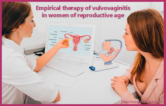
Empirical therapy of vulvovaginitis in women

Empirical therapy of vulvovaginitis in women: At the turn of the millennium, a real revolution took place in our understanding of vaginal infections. The Koch postulate “one disease-one pathogen”, which was once accepted as an axiom, was replaced by the understanding that in the vast majority of cases these conditions have a mixed etiology, and nosological forms caused by a single pathogen are rare. When treating a patient with bacterial vaginosis or vaginitis, the obstetrician-gynecologist should be guided by new data on the causes and pathogenesis of these diseases. We won’t talk about clomid today.

“Lonely” microbes are rare, usually there is a “cocktail” of bacteria, fungi, viruses and protozoa. A wide variety of” permanent inhabitants “of the biotope ensures its normal functional state, and also prevents colonization by” external ” obligate and facultative pathogens of diseases. In one patient, mutually exclusive states of the vaginal microbiome can be observed simultaneously. For example, candidiasis or aerobic vaginitis, the” instigators ” of which need oxygen, and bacterial vaginosis caused by anaerobic microorganisms.

J. Sobel distinguishes two types of mixed infections: mixed infection and co-infection. In mixed infections, all pathogens jointly damage the mucous membrane of the genital tract, entering into complex relationships aimed at increasing virulence and drug resistance. Coinfections are detected against the background of an active inflammatory process caused by a dominant pathogen. The rest remain invisible and manifest themselves only after the elimination of the predecessor.

Antibiotic resistance is an interdisciplinary and inter-state problem, for the effective solution of which, both at the hospital and outpatient level, it is necessary to consolidate doctors of different specialties. The growing resistance of microorganisms to antibiotics poses a difficult task for the obstetrician-gynecologist. The scientific justification for the combination of most vaginal infections and vaginal dysbiosis makes doctors more scrupulous in their diagnosis and treatment. A drug for the local therapy of vulvovaginitis should “cover” the entire spectrum of possible pathogens due to its constituent substances of multidirectional action. In order to avoid reducing compliance, instead of several drugs, it is advisable to prescribe a combined drug with the widest possible spectrum of action, covering most of the potential pathogens of vaginal infection – bacteria, fungi and protozoa.

Empirical therapy of vulvovaginitis: Materials and methods

Exclusion criteria:

  • hypersensitivity to one or more components of the drug Gainomax;
  • taking antiseptics, antibiotics, and antibiotics at the time of inclusion in the study;
  • the need for systemic use of glucocorticosteroids, cytostatics and systemic antibiotics;
  • documented HIV infection, syphilis and other sexually transmitted infections (chlamydia infection, gonorrhea, trichomoniasis) detected during the screening stage;
  • acute somatic diseases;
  • the period of pregnancy or breastfeeding.

Patient follow-up included consecutive visits.

At the first visit (screening, inclusion in the study), the written informed consent of the patients was obtained, the inclusion criteria were checked, and the exclusion criteria were evaluated. After examination and taking the material for analysis (bacterioscopy and pH-metry of vaginal secretions, real-time polymerase chain reaction (PCR) analysis (Femoflor-16)), based on complaints and clinical symptoms, the patients were immediately prescribed empirical therapy with the complex antimicrobial drug Gainomax. All patients were divided into two representative groups. 36 (50%) patients of the first group took Gainomax one suppository once a day for seven days, according to the approved instructions; 36 (50%) patients of the second group – Gainomax one suppository twice a day for three days, according to the approved instructions.

Empirical therapy of vulvovaginitis

At the second visit, 10-12 days after the completion of seven – or three-day therapy with Gainomax, a control examination was performed, which included a repeated gynecological examination, an assessment of the dynamics of clinical symptoms, the pH index and the microbial landscape of the vaginal discharge. Satisfaction and compliance of therapy, adverse events associated with the use of Gainomax were evaluated, the proportion of patients with a complete cure was determined, and cases of ineffectiveness of therapy were recorded. All medical procedures performed in the study were routine, used in everyday clinical practice, which determined the non-interventional (observational) nature of the study.

Conclusion

The analysis of the effectiveness of empirical therapy of acute vulvovaginitis using the drug Gainomax showed no complaints of abnormal vaginal discharge at the second visit in 94.4% of patients of the first and 97.2% of patients of the second group. A significant decrease in the volume of vaginal discharge was noted by 83.3% and 80.6% of patients of the first and second groups, respectively. The complete disappearance of unpleasant odors, itching and burning sensations in the vagina was indicated by 91.7% of the patients of the first group and 94.4% of the patients of the second group. When objectively evaluated, normalization of the color of the vaginal walls, moderate volume and “slimy” nature of secretions were recorded in all participants of the study. In most of them, bacterioscopy of vaginal smears after empirical therapy revealed a significant decrease in the number of white blood cells in the visual field. After therapy, yeast-like fungi of the genus Candida were not detected in any patient.

Modern view on the problem of tubal-peritoneal infertility

Tubal-peritoneal factor is currently the leading cause of female infertility, accounting, according to various authors, from 20 to 72%. Despite the achievements of modern medicine, tubal-peritoneal infertility is still one of the most serious pathological conditions, taking into account the difficulty of its diagnosis and treatment, as well as the possibility of restoring reproductive function.

The main causes of tubal-peritoneal infertility are:

  • previously transmitted inflammatory diseases of the pelvic organs of a specific and non-specific nature, of which the most important are chlamydia, gonorrhea, mycoplasma and trichomonas infections;
  • various intrauterine manipulations (artificial termination of pregnancy, separate diagnostic scraping of the endometrium and endocervix, hysteroscopy with removal of endometrial polyps or submucous myomatous nodes, etc.);
  • postpartum and postabortem complications of traumatic and inflammatory genesis;
  • previous surgical interventions on the pelvic organs (ovarian resection, conservative myomectomy, tubectomy, tubal ligation) and the abdominal cavity, especially those performed according to emergency indications from traditional laparotomy access and complicated by the development of peritonitis (for example, appendectomy).

Damage to the abdominal cavity during surgery and the subsequent development of aseptic inflammation lead to the deposition of fibrin in the area of the surgical wound with local activation of fibrinolysis and proteolysis, which ultimately contributes to the resorption of primary fibrinous deposits without the formation of adhesions. With the development of postoperative infection, the inflammatory-dystrophic process is delayed, which does not allow fibrinous formations to dissolve and contributes to excessive local collagen production with the formation of powerful connective tissue junctions – adhesions.

tubal-peritoneal infertility

The severity of the adhesive process in the pelvis directly depends on the extent of the spread of adhesions in the abdominal cavity, which is mainly determined by the volume and type of surgical intervention. In this regard, in order to prevent the development of the adhesive process in the abdominal cavity, one should strive to minimize the surgical intervention, giving preference to the laparoscopic method (especially when performing planned surgical interventions on the pelvic organs in women of reproductive age).

Treatment of tubal-peritoneal infertility

Treatment of tubal-peritoneal infertility includes conservative and surgical methods used sequentially or in combination with each other. Conservative treatment of tubal-peritoneal infertility involves anti-inflammatory antibacterial, immunomodulatory, antifibrosing therapy and physiotherapy.

When sexually transmitted infections are detected and / or a morphologically verified diagnosis of chronic endometritis, therapy should be comprehensive, etiopathogenetic and aimed at the complete elimination of the identified pathogens. Immunomodulatory therapy is an indispensable part of the treatment of tubal-peritoneal infertility, since chronic inflammatory processes of the pelvic organs are always accompanied by immunological disorders, which requires mandatory correction.

One of the options for antifibrosing therapy is the general and local use of various drugs with a resorbing effect – biostimulants, enzymes and glucocorticosteroids (hydrocortisone) in the form of tampons, as well as hydrotubation. Unfortunately, the clinical experience of using hydrotubation as a method of local treatment of tubal-peritoneal infertility has demonstrated insufficient effectiveness and a high frequency of various complications. Among them, most often there is an exacerbation of chronic inflammatory diseases of the pelvic organs with a violation of the functional ability of endosalpinx cells and the development of hydrosalpinx, which significantly reduces the peristaltic activity of the fallopian tubes and disrupts the transport of gametes through them.

Thus, tubal-peritoneal infertility requires early diagnosis and gradual long-term rehabilitation after undergoing surgical treatment. Also remember that ovulation stimulation is possible with the help of the drug clomid. One of the most effective ways to preserve the reproductive function is the prevention of tubal-peritoneal infertility, which consists in the prevention and timely treatment of inflammatory diseases of the pelvic organs, the rational management of childbirth and the postpartum period, and the implementation of rehabilitation measures in the near future after gynecological operations.